Category: Uncategorized

Two green-stained cross sections of a mouse brain, one labeled Cuprizone Control and one labeled Cuprizone 40Hz,, are accompanied by insets showing magnifications of parts specific brain regions. The green staining in each inset is greater and brighter in the 40Hz insets vs. the control insets.

Study reveals ways in which 40Hz sensory stimulation may preserve brain’s ‘white matter’

MIT scientists report that gamma frequency light and sound stimulation preserves myelination in mouse models and reveal molecular mechanisms that may underlie the benefit.

Early-stage trials in Alzheimer’s disease patients and studies in mouse models of the disease have suggested positive impacts on pathology and symptoms from exposure to light and sound presented at the “gamma” band frequency of 40 Hz. A new study zeroes in on how 40Hz sensory stimulation helps to sustain an essential process in which the signal-sending branches of neurons, called axons, are wrapped in a fatty insulation called myelin. Often called the brain’s “white matter,” myelin protects axons and insures better electrical signal transmission in brain circuits.

“Previous publications from our lab have mainly focused on neuronal protection,” said Li-Huei Tsai, Picower Professor in The Picower Institute for Learning and Memory and the Department of Brain and Cognitive Sciences at MIT and senior author of the new study in Nature Communications. Tsai also lead’s MIT’s Aging Brain Initiative. “But this study shows that it’s not just the gray matter, but also the white matter that’s protected by this method.”

This year Cognito Therapeutics, the spin-off company that licensed MIT’s sensory stimulation technology, published phase II human trial results in the Journal of Alzheimer’s Disease indicating that 40Hz light and sound stimulation significantly slowed the loss of myelin in volunteers with Alzheimer’s. Also this year Tsai’s lab published a study showing that gamma sensory stimulation helped mice withstand neurological effects of chemotherapy medicines, including by preserving myelin. In the new study, members of Tsai’s lab led by former postdoc Daniela Rodrigues Amorim used a common mouse model of myelin loss—a diet with the chemical cuprizone— to explore how sensory stimulation preserves myelination.

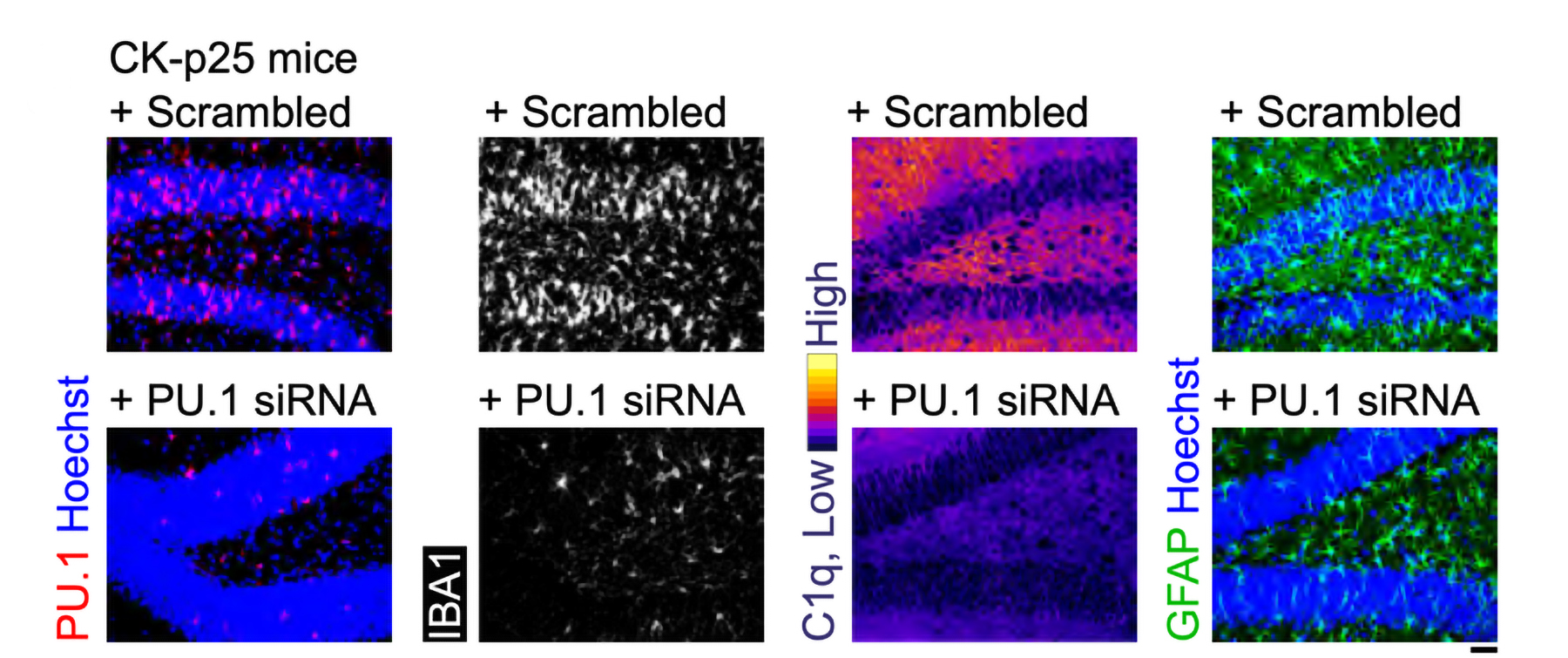
Amorim and Tsai’s team found that 40Hz light and sound not only preserved myelination in the brains of cuprizone-exposed mice, it also appeared to protect oligodendrocytes (the cells that myelinate neural axons), sustain the electrical performance of neurons, and preserve a key marker of axon structural integrity. When the team looked into the molecular underpinnings of these benefits, they found clear signs of specific mechanisms including preservation of neural circuit connections called synapses; a reduction in a cause of oligodendrocyte death called “ferroptosis;” reduced inflammation; and an increase in the ability of microglia brain cells to clean up myelin damage so that new myelin could be restored.

“Gamma stimulation promotes a healthy environment,” said Amorim who is now a Marie Curie Fellow at the University of Galway in Ireland. “There are several ways we are seeing different effects.”

The findings suggest that gamma sensory stimulation may help not only Alzheimer’s disease patients but also people battling other diseases involving myelin loss, such as multiple sclerosis, the authors wrote in the study.

Maintaining myelin

To conduct the study, Tsai and Amorim’s team fed some male mice a diet with cuprizone and gave other male mice a normal diet for six weeks. Halfway into that period, when cuprizone is known to begin causing its most acute effects on myelination, they exposed some mice from each group to gamma sensory stimulation for the remaining three weeks. In this way they had four groups: completely unaffected mice, mice that received no cuprizone but did get gamma stimulation, mice that received cuprizone and constant (but not 40Hz) light and sound as a control, and mice that received cuprizone and also gamma stimulation.

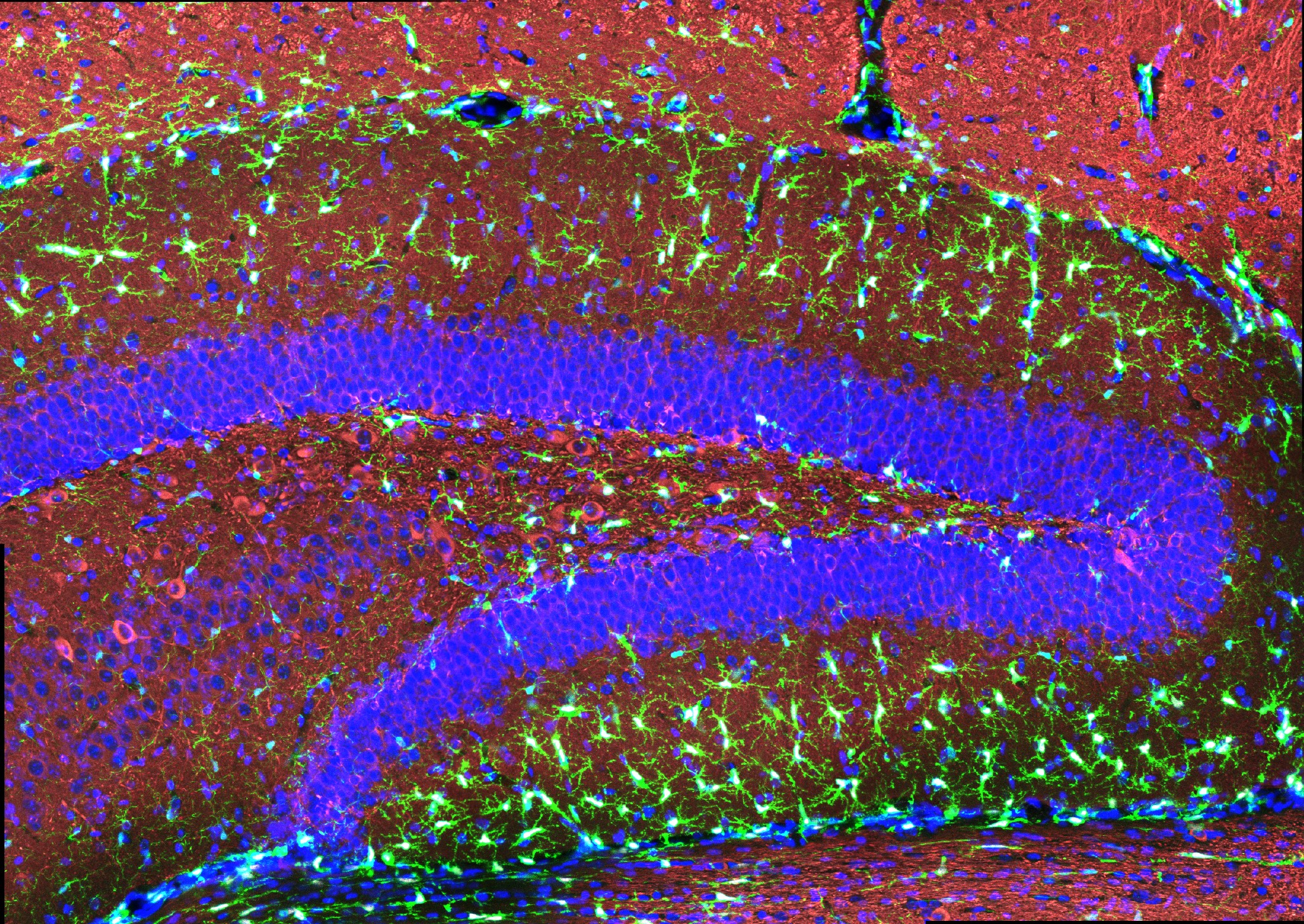
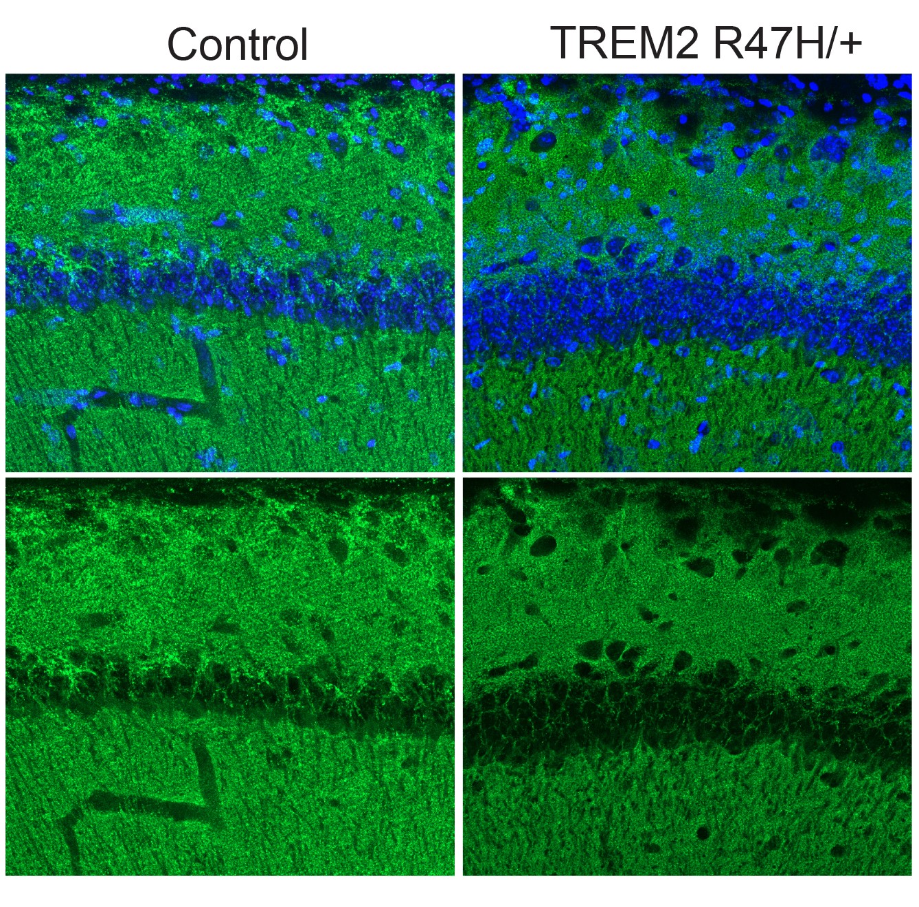
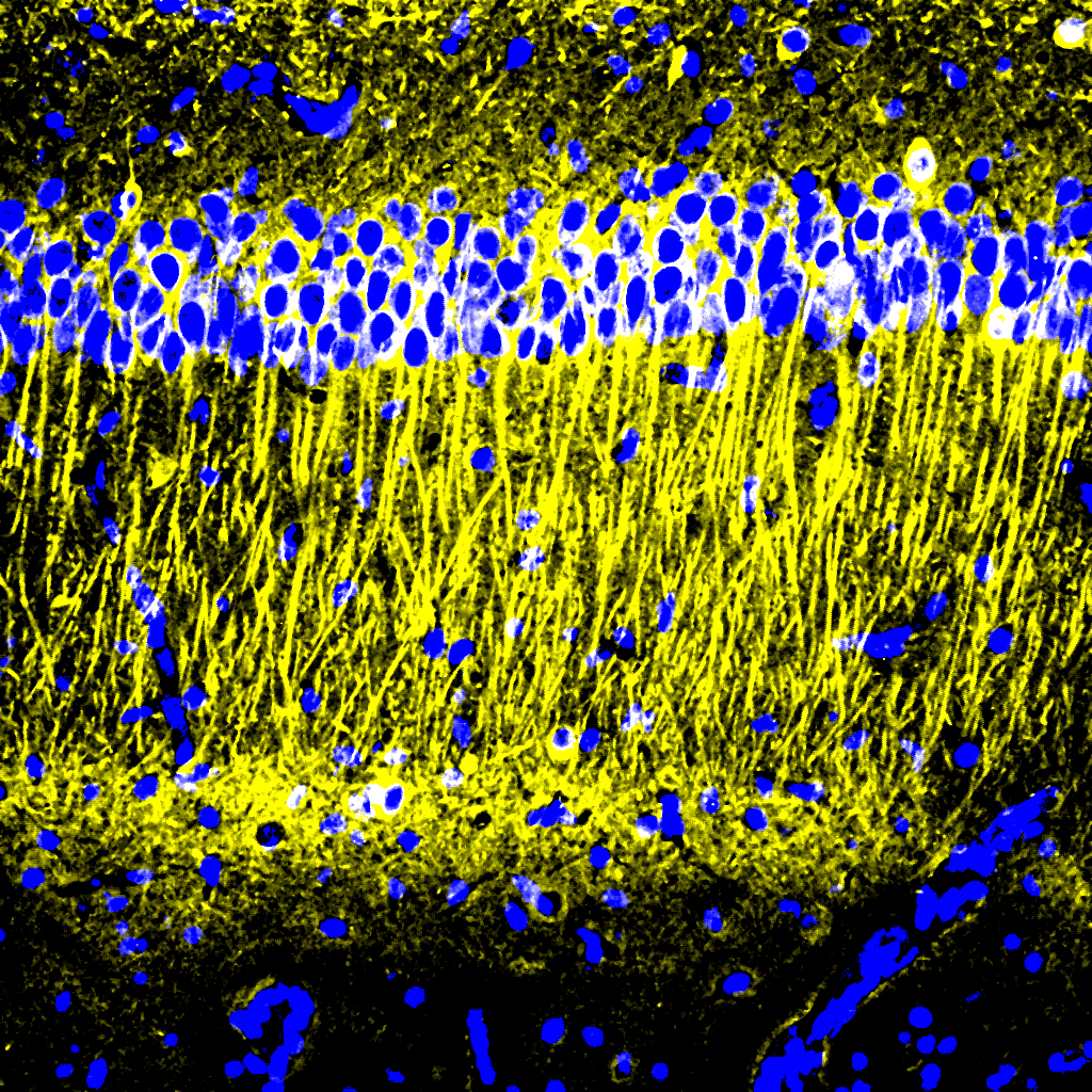
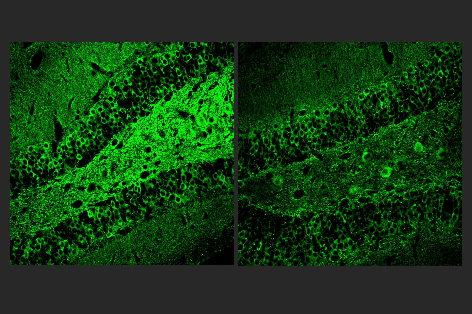
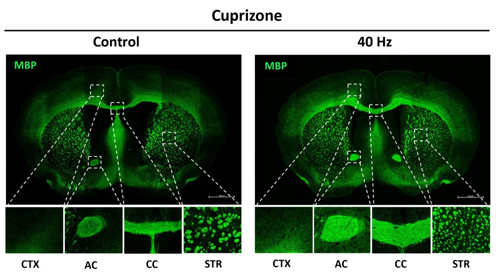
After the six weeks elapsed, the scientists measured signs of myelination throughout the brains of the mice in each group. Mice that weren’t fed cuprizone maintained healthy levels, as expected. Mice that were fed cuprizone and didn’t receive 40Hz gamma sensory stimulation showed drastic levels of myelin loss. Cuprizone-fed mice that received 40Hz stimulation retained significantly more myelin, rivaling the health of mice never fed cuprizone by some, but not all, measures.

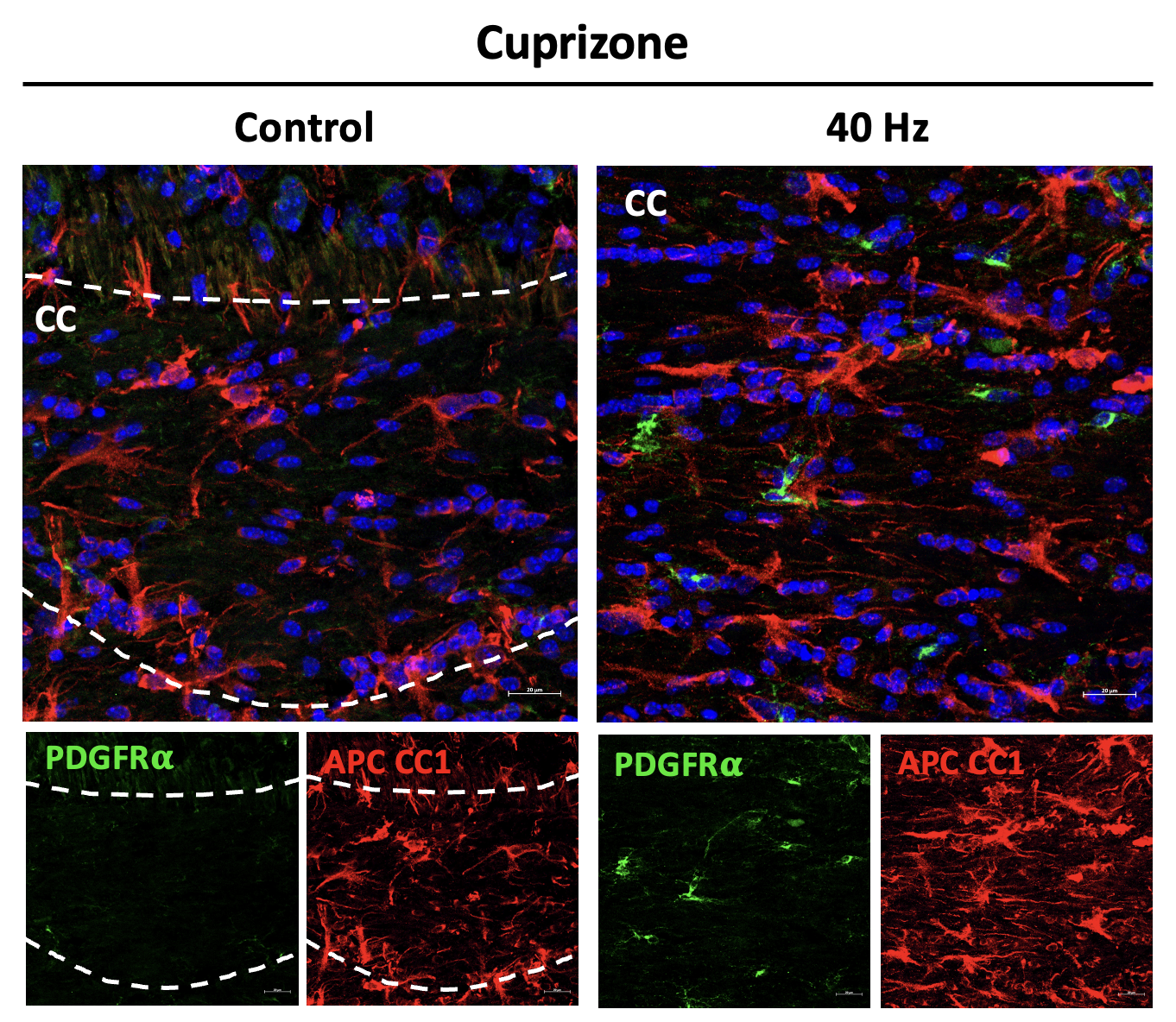
The researchers also looked at numbers of oligodendrocytes to see if they survived better with sensory stimulation. Several measures revealed that in mice fed cuprizone, oligodendrocytes in the corpus callosum region of the brain (a key point for the transit of neural signals because it connects the brain’s hemispheres) were markedly reduced. But in mice fed cuprizone and also treated with gamma stimulation, the number of cells were much closer to healthy levels.

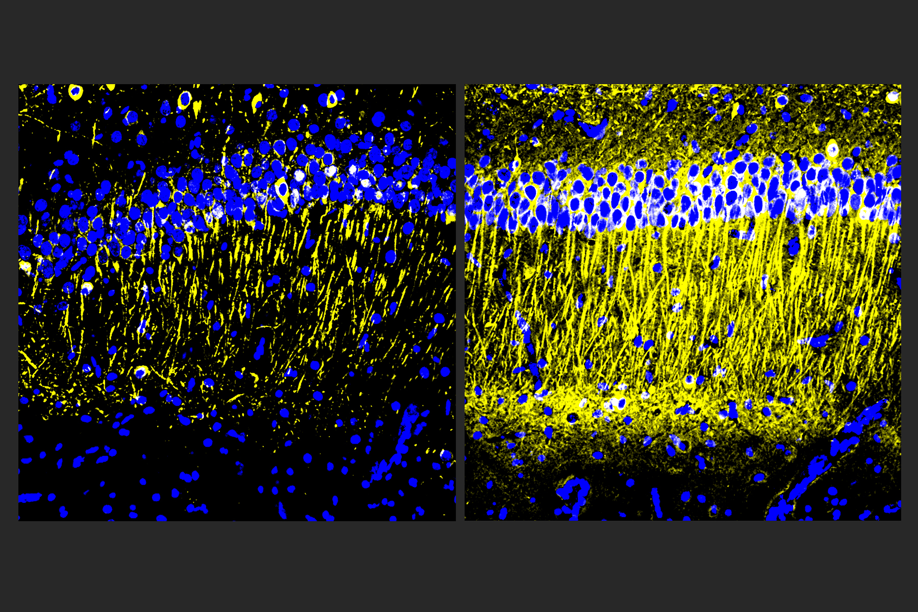
Electrophysiological tests among neural axons in the corpus callosum showed that gamma sensory stimulation was associated with improved electrical performance in cuprizone-fed mice who received gamma stimulation compared to cuprizone-fed mice left untreated by 40Hz stimulation. And when researchers looked in the anterior cingulate cortex region of the brain, they saw that MAP2, a protein that signals the structural integrity of axons, was much better preserved in mice that received cuprizone and gamma stimulation compared to cuprizone-fed mice who did not.

Side by side panels labeled cuprizone control and cuprizone 40hz compare amounts of cells stained red and green. The 40Hz panel shows much more red and green staining.
To assess the number of immature and mature oligodendrocytes in the corpus callosum in mice fed cuprizone and given 40 Hz stimulation (right), compared to mice fed cuprizone and given constant stimulation as a control, researchers measured the marker PDGFRa (green) and APCCC1 (red). The 40Hz group exhibited a signficantly higher number of each type of oligodendrocytes.

Molecular mechanisms

A key goal of the study was to identify possible ways in which 40Hz sensory stimulation may protect myelin.

To find out, the researchers conducted a sweeping assessment of protein expression in each mouse group and identified which proteins were differentially expressed based on cuprizone diet and exposure to gamma frequency stimulation. The analysis revealed distinct sets of effects between the cuprizone mice exposed to control stimulation and cuprizone-plus-gamma mice.

A highlight of one set of effects was the increase in MAP2 in gamma-treated cuprizone-fed mice. A highlight of another set was that cuprizone mice who received control stimulation showed a substantial deficit in expression of proteins associated with synapses. The gamma-treated cuprizone-fed mice did not show any significant loss, mirroring results in a 2019 Alzheimer’s 40Hz study that showed synaptic preservation. This result is important, the researchers wrote, because neural circuit activity, which depends on maintaining synapses, is associated with preserving myelin. They confirmed the protein expression results by looking directly at brain tissues.

Another set of protein expression results hinted at another important mechanism: ferroptosis. This phenomenon, in which errant metabolism of iron leads to a lethal buildup of reactive oxygen species in cells, is a known problem for oligodendrocytes in the cuprizone mouse model. Among the signs was an increase in cuprizone-fed, control stimulation mice in expression of the protein HMGB1, which is a marker of ferroptosis-associated damage that triggers an inflammatory response. Gamma stimulation, however, reduced levels of HMGB1.

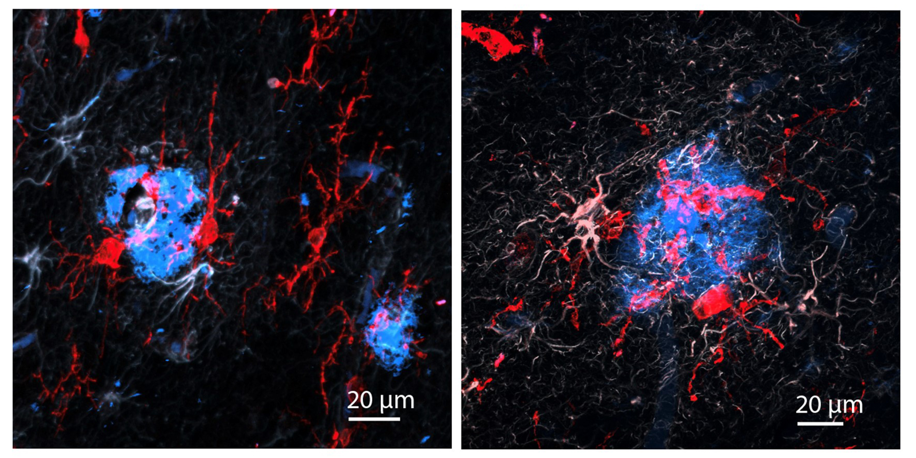
Looking more deeply at the cellular and molecular response to cuprizone demyelination and the effects of gamma stimulation, the team assessed gene expression using single-cell RNA sequencing technology. They found that astrocytes and microglia became very inflammatory in cuprizone-control mice but gamma stimulation calmed that response. Fewer cells became inflammatory and direct observations of tissue showed that microglia became more proficient at clearing away myelin debris, a key step in effecting repairs.

The team also learned more about how oligodendrocytes in cuprizone-fed mice exposed to 40Hz sensory stimulation managed to survive better. Expression of protective proteins such as HSP70 increased and as did expression of GPX4, a master regulator of processes that constrain ferroptosis.

In addition to Amorim and Tsai, the paper’s other authors are Lorenzo Bozzelli, TaeHyun Kim, Liwang Liu, Oliver Gibson, Cheng-Yi Yang, Mitch Murdock, Fabiola Galiana-Meléndez, Brooke Schatz, Alexis Davison, Md Rezaul Islam, Dong Shin Park, Ravikiran M. Raju, Fatema Abdurrob, Alissa J. Nelson, Jian Min Ren, Vicky Yang and Matthew P. Stokes.

Fundacion Bancaria la Caixa, The JPB Foundation, The Picower Institute for Learning and Memory, the Carol and Gene Ludwig Family Foundation, Lester A. Gimpelson, Eduardo Eurnekian, The Dolby Family, Kathy and Miguel Octavio, the Marc Haas Foundation, Ben Lenail and Laurie Yoler, and the National Institutes of Health provided funding for the study.

Study across multiple brain regions discerns Alzheimer’s vulnerability and resilience factors

Genomics and lab studies reveal numerous findings, including a key role for Reelin amid neuronal vulnerability, and for choline and antioxidants in sustaining cognition

An MIT study published today in Nature provides new evidence for how specific cells and circuits become vulnerable in Alzheimer’s disease, and hones in on other factors that may help some people show resilience to cognitive decline, even amid clear signs of disease pathology. To highlight potential targets for interventions to sustain cognition and memory, the authors engaged in a novel comparison of gene expression across multiple brain regions in people with or without Alzheimer’s disease, and conducted lab experiments to test and validate their major findings.

Brain cells all have the same DNA but what makes them differ, both in their identity and their activity, are their patterns of how they express those genes. The new analysis measured gene expression differences in more than 1.3 million cells of more than 70 cell types in six brain regions from 48 tissue donors, 26 of whom died with an Alzheimer’s diagnosis and 22 of whom without. As such, the study provides a uniquely large, far-ranging and yet detailed accounting of how brain cell activity differs amid Alzheimer’s disease by cell type, by brain region, by disease pathology, and by each person’s cognitive assessment while still alive.

“Specific brain regions are vulnerable in Alzheimer’s and there is an important need to understand how these regions or particular cell types are vulnerable,” said co-senior author Li-Huei Tsai, Picower Professor of Neuroscience and director of The Picower Institute for Learning and Memory and the Aging Brain Initiative at MIT. “And the brain is not just neurons. It’s many other cell types. How these cell types may respond differently, depending on where they are, is something fascinating we are only at the beginning of looking at.”

Co-senior author Manolis Kellis, professor of computer science and head of MIT’s Computational Biology Group, likened the technique used to measure gene expression comparisons, single cell RNA profiling, to being a much more advanced “microscope” than the ones that first allowed Alois Alzheimer to characterize the disease’s pathology more than a century ago.

“Where Alzheimer saw amyloid protein plaques and phosphorylated tau tangles in his microscope, our single-cell ‘microscope’ tells us, cell by cell and gene by gene, about thousands of subtle yet important biological changes in response to pathology,” said Kellis. “Connecting this information with the cognitive state of patients reveals how cellular responses relate with cognitive loss or resilience, and can help propose new ways to treat cognitive loss. Pathology can precede cognitive symptoms by a decade or two before cognitive decline becomes diagnosed. If there’s not much we can do about the pathology at that stage, we can at least try to safeguard the cellular pathways that maintain cognitive function.”

Hansruedi Mathys, a former MIT postdoc in the Tsai Lab, who is now an assistant professor at the University of Pittsburgh, Carles Boix, a former graduate student in Kellis’s lab who is now a postdoc at Harvard Medical School, and Leyla Akay, a graduate student in Tsai’s lab, led the study analyzing the prefrontal cortex, entorhinal cortex, hippocampus, anterior thalamus, angular gyrus, and the midtemporal cortex. The brain samples came from the Religious Order Study and the Rush Memory and Aging Project at Rush University.

Neural vulnerability and Reelin

Some of the earliest signs of amyloid pathology and neuron loss in Alzheimer’s occurs in memory-focused regions called the hippocampus and the entorhinal cortex. In those regions, and in other parts of the cerebral cortex, the researchers were able to pinpoint a potential reason why. One type of excitatory neuron in the hippocampus and four in the entorhinal cortex were significantly less abundant in people with Alzheimer’s than in people without. Individuals with depletion of those cells performed significantly worse on cognitive assessments. Moreover, many vulnerable neurons were interconnected in a common neuronal circuit. And just as importantly, several either directly expressed a protein called Reelin, or were directly affected by Reelin signaling. In all, therefore, the findings distinctly highlight especially vulnerable neurons, whose loss is associated with reduced cognition, that share a neuronal circuit and a molecular pathway.

Tsai noted that Reelin has become prominent in Alzheimer’s research because of a recent study of a man in Colombia. He had a rare mutation in the Reelin gene that caused the protein to be more active, and was able to stay cognitively healthy at an advanced age despite having a strong family predisposition to early-onset Alzheimer’s. The new study shows that loss of Reelin-producing neurons is associated with cognitive decline. Taken together it may mean that the brain benefits from Reelin, but that neurons that produce it may be lost in at least some Alzheimer’s patients.

“We can think of Reelin as having maybe some kind of protective or beneficial effect,” Akay said. “But we don’t yet know what it does or how it could confer resilience.”

In further analysis the researchers also found that specifically vulnerable inhibitory neuron subtypes identified in a previously study from this group in the prefrontal cortex also were involved in reelin signaling, further reinforcing the significance of the molecule and its signaling pathway.

To further check their results, the team directly examined the human brain tissue samples and the brains of two kinds of Alzheimer’s model mice. Sure enough, those experiments also showed a reduction in Reelin-positive neurons in the human and mouse entorhinal cortex.

Resilience associated with choline metabolism in astrocytes

To find factors that might preserve cognition, even amid pathology, the team examined which genes, in which cells, and in which regions, were most closely associated with cognitive resilience, which they defined as residual cognitive function, above the typical cognitive loss expected given the observed pathology.

Their analysis yielded a surprising and specific answer: across several brain regions astrocytes that expressed genes associated with antioxidant activity and with choline metabolism and polyamine biosynthesis were significantly associated with sustained cognition, even amid high levels of tau and amyloid. The results reinforced previous research findings led by Tsai and Susan Lundqvist in which they showed that dietary supplement of choline helped astrocytes cope with the dysregulation of lipids caused by the most significant Alzheimer’s risk gene, the APOE4 variant. The antioxidant findings also pointed to a molecule that can be found as a dietary supplement, spermidine, which may have anti-inflammatory properties, although such an association would need further work to be established causally.

As before, the team went beyond the predictions from the single-cell RNA expression analysis to make direct observations in the brain tissue of samples. Those that came from cognitively resilient individuals indeed showed increased expression of several of the astrocyte-expressed genes predicted to be associated with cognitive resilience.

In two columns labeled "Cognitively Impaired" and "Cognitively Resistent" microscope images show astrocyte cells with magenta staining. Small blue dots are visible around some of the astrocyte cells and those dots are indicated by large white arrows.
Expression of the gene GPCPD1 in astrocyte cells is associated with cognitive resilience in people with Alzheimer’s pathology. Here white arrows indicate instances of GPCPD1 expression (blue) in astrocyte cells (denoted by AQP4 staining in magenta). There is much more expression in tissue from the cognitively resilient person (right).

New analysis method, open dataset

To analyze the mountains of single-cell data, the researchers developed a new robust methodology based on groups of coordinately-expressed genes (known as “gene modules”), thus exploiting the expression correlation patterns between functionally-related genes in the same module.

“In principle, the 1.3 million cells we surveyed could use their 20,000 genes in an astronomical number of different combinations,” explain Kellis. “In practice, however, we observe a much smaller subset of coordinated changes. Recognizing these coordinated patterns allow us to infer much more robust changes, because they are based on multiple genes in the same functionally-connected module.”

He offered this analogy: With many joints in their bodies, people could move in all kinds of crazy ways, but in practice they engage in many fewer coordinated movements like walking, running, or dancing. The new method enables scientists to identify such coordinated gene expression programs as a group.

While Kellis and Tsai’s labs already reported several noteworthy findings from the dataset, the researchers expect that many more possibly significant discoveries still wait to be found in the trove of data. To facilitate such discovery the team posted handy analytical and visualization tools along with the data on Kellis’s website at: https://compbio-mit-edu.ezproxy.canberra.edu.au/ad_multiregion.

“The dataset is so immensely rich. We focused on only a few aspects that are salient that we believe are very, very interesting, but by no means have we exhausted what can be learned with this dataset,” Kellis said. “We expect many more discoveries ahead, and we hope that young researchers (of all ages) will dive right in and surprise us with many more insights.”

Going forward, Kellis said, the researchers are studying the control circuitry associated with the differentially expressed genes, to understand the genetic variants, the regulators, and other driver factors that can be modulated to reverse disease circuitry across brain regions, cell types, and different stages of the disease.

Additional authors of the study include Ziting Xia, Jose Davila Velderrain, Ayesha P. Ng, Xueqiao Jiang, Ghada Abdelhady, Kyriaki Galani, Julio Mantero, Neil Band, Benjamin T. James, Sudhagar Babu, Fabiola Galiana-Melendez, Kate Louderback, Dmitry Prokopenko, Rudolph E. Tanzi, and David A. Bennett.

Support for the research came from the National Institutes of Health, The Picower Institute for Learning and Memory, The JPB Foundation, the Cure Alzheimer’s Fund, The Robert A. and Renee E. Belfer Family Foundation, Eduardo Eurnekian, and Joseph DiSabato.

Two panels on a black background show cells on the left lit brightly in green while the cells on the right show much less green and are mostly red.

A noninvasive treatment for “chemo brain”

Stimulating gamma brain waves may protect cancer patients from memory impairment and other cognitive effects of chemotherapy.

Patients undergoing chemotherapy often experience cognitive effects such as memory impairment and difficulty concentrating — a condition commonly known as “chemo brain.”

MIT researchers have now shown that a noninvasive treatment that stimulates gamma frequency brain waves may hold promise for treating chemo brain. In a study of mice, they found that daily exposure to light and sound with a frequency of 40 hertz protected brain cells from chemotherapy-induced damage. The treatment also helped to prevent memory loss and impairment of other cognitive functions.

This treatment, which was originally developed as a way to treat Alzheimer’s disease, appears to have widespread effects that could help with a variety of neurological disorders, the researchers say.

“The treatment can reduce DNA damage, reduce inflammation, and increase the number of oligodendrocytes, which are the cells that produce myelin surrounding the axons,” says Li-Huei Tsai, director of MIT’s Picower Institute for Learning and Memory and Picower Professor in the MIT Department of Brain and Cognitive Sciences. “We also found that this treatment improved learning and memory, and enhanced executive function in the animals.”

Tsai, who also leads MIT’s Aging Brain Initiative, is the senior author of the new study, which appears today in Science Translational Medicine. The paper’s lead author is TaeHyun Kim, an MIT postdoc.

Protective brain waves

Several years ago, Tsai and her colleagues began exploring the use of light flickering at 40 hertz (cycles per second) as a way to improve the cognitive symptoms of Alzheimer’s disease. Previous work had suggested that Alzheimer’s patients have impaired gamma oscillations — brain waves that range from 25 to 80 hertz (cycles per second) and are believed to contribute to brain functions such as attention, perception, and memory.

Tsai’s studies in mice have found that exposure to light flickering at 40 hertz or sounds with a pitch of 40 hertz can stimulate gamma waves in the brain, which has many protective effects, including preventing the formation of amyloid beta plaques. Using light and sound together provides even more significant protection. The treatment also appears promising in humans: Phase 1 clinical trials in people with early-stage Alzheimer’s disease have found the treatment is safe and does offer some neurological and behavioral benefits.

In the new study, the researchers set out to see whether this treatment could also counteract the cognitive effects of chemotherapy treatment. Research has shown that these drugs can induce inflammation in the brain, as well as other detrimental effects such as loss of white matter — the networks of nerve fibers that help different parts of the brain communicate with each other. Chemotherapy drugs also promote loss of myelin, the protective fatty coating that allows neurons to propagate electrical signals. Many of these effects are also seen in the brains of people with Alzheimer’s.

“Chemo brain caught our attention because it is extremely common, and there is quite a lot of research on what the brain is like following chemotherapy treatment,” Tsai says. “From our previous work, we know that this gamma sensory stimulation has anti-inflammatory effects, so we decided to use the chemo brain model to test whether sensory gamma stimulation can be beneficial.”

On a black background two panels show spiny microglia cells. In the left panel many are bright red but in the right panel there is very little red coloration
Red staining denotes microglia brain cells bearing the microglia marker Iba1 and the green staining microglia inflammatory marker CD68. In mice exposed to cisplatin and left untreated by 40Hz stimulation (left), a lot of red and green staining is evident. In cisplatin-exposed mice who received 40Hz treatment (right) the red and green staining (and inflammation) is reduced.

As an experimental model, the researchers used mice that were given cisplatin, a chemotherapy drug often used to treat testicular, ovarian, and other cancers. The mice were given cisplatin for five days, then taken off of it for five days, then on again for five days. One group received chemotherapy only, while another group was also given 40-hertz light and sound therapy every day.

After three weeks, mice that received cisplatin but not gamma therapy showed many of the expected effects of chemotherapy: brain volume shrinkage, DNA damage, demyelination, and inflammation. These mice also had reduced populations of oligodendrocytes, the brain cells responsible for producing myelin.

However, mice that received gamma therapy along with cisplatin treatment showed significant reductions in all of those symptoms. The gamma therapy also had beneficial effects on behavior: Mice that received the therapy performed much better on tests designed to measure memory and executive function.

“A fundamental mechanism”

Using single-cell RNA sequencing, the researchers analyzed the gene expression changes that occurred in mice that received the gamma treatment. They found that in those mice, inflammation-linked genes and genes that trigger cell death were suppressed, especially in oligodendrocytes, the cells responsible for producing myelin.

In mice that received gamma treatment along with cisplatin, some of the beneficial effects could still be seen up to four months later. However, the gamma treatment was much less effective if it was started three months after the chemotherapy ended.

The researchers also showed that the gamma treatment improved the signs of chemo brain in mice that received a different chemotherapy drug, methotrexate, which is used to treat breast, lung, and other types of cancer.

“I think this is a very fundamental mechanism to improve myelination and to promote the integrity of oligodendrocytes. It seems that it’s not specific to the agent that induces demyelination, be it chemotherapy or another source of demyelination,” Tsai says.

Because of its widespread effects, Tsai’s lab is also testing gamma treatment in mouse models of other neurological diseases, including Parkinson’s disease and multiple sclerosis. Cognito Therapeutics, a company founded by Tsai and MIT Professor Edward Boyden, has finished a phase 2 trial of gamma therapy in Alzheimer’s patients, and plans to begin a phase 3 trial this year.

“My lab’s major focus now, in terms of clinical application, is Alzheimer’s; but hopefully we can test this approach for a few other indications, too,” Tsai says.

The research was funded by the JPB Foundation, the Ko Hahn Seed Fund, and the National Institutes of Health.

–From MIT News

A coronal cross-section of a mouse brain is stained blue. The entire outer edge and occasional points further inside are speckled with yellow-green dots. The background behind the brain is black.

How sensory gamma rhythm stimulation clears amyloid in Alzheimer’s mice

Stimulating a key brain rhythm with light and sound increases peptide release from interneurons, driving clearance of Alzheimer’s protein via the brain’s glymphatic system, new study suggests.

Studies at MIT and elsewhere are producing mounting evidence that light flickering and sound clicking at the gamma brain rhythm frequency of 40 Hz can reduce Alzheimer’s disease (AD) progression and treat symptoms in human volunteers as well as lab mice. In a new study in Nature using a mouse model of the disease, researchers at The Picower Institute for Learning and Memory of MIT reveal a key mechanism that may contribute to these beneficial effects: clearance of amyloid proteins, a hallmark of AD pathology, via the brain’s glymphatic system, a recently discovered “plumbing” network parallel to the brain’s blood vessels.

“Ever since we published our first results in 2016, people have asked me how does it work? Why 40 Hz? Why not some other frequency?” said study senior author Li-Huei Tsai, Picower Professor of Neuroscience and director of The Picower Institute and MIT’s Aging Brain Initiative. “These are indeed very important questions we have worked very hard in the lab to address.”

The new paper describes a series of experiments, led by Mitch Murdock when he was a Brain and Cognitive Sciences doctoral student at MIT, showing that when sensory gamma stimulation increases 40 Hz power and synchrony in the brains of mice, that prompts a particular type of neuron to release peptides. The study results further suggest that those short protein signals then drive specific processes that promote increased amyloid clearance via the glymphatic system.

“We do not yet have a linear map of the exact sequence of events that occurs,” said Murdock, who was jointly supervised by Tsai and co-author and collaborator Ed Boyden, Y. Eva Tan Professor of Neurotechnology at MIT, a member of the McGovern Institute for Brain Research and an affiliate member of The Picower Institute. “But the findings in our experiments support this clearance pathway through the major glymphatic routes.”

From Gamma to Glymphatics

Because prior research has shown that the glymphatic system is a key conduit for brain waste clearance and may be regulated by brain rhythms, Tsai and Murdock’s team hypothesized that it might help explain the lab’s prior observations that gamma sensory stimulation reduces amyloid levels in Alzheimer’s model mice.

Working with “5XFAD” mice, which genentically model Alzheimer’s, Murdock and co-authors  first replicated the lab’s prior results that 40 Hz sensory stimulation increases 40 Hz neuronal activity in the brain and reduces amyloid levels. Then they set out to measure whether there was any correlated change in the fluids that flow through the glymphatic system to carry away wastes. Indeed, they measured increases in cerebrospinal fluid  in the brain tissue of mice treated with sensory gamma stimulation compared to untreated controls. They also measured an increase in the rate of interstitial fluid leaving the brain. Moreover, in the gamma-treated mice he measured increased diameter of the lymphatic vessels that drain away the fluids and measured increased accumulation of amyloid in cervical lymph nodes, which is the drainage site for that flow.

To investigate how this increased fluid flow might be happening, the team focused on the aquaporin 4 (AQP4) water channel of astrocyte cells, which enables the cells to facilitate glymphatic fluid exchange. When they blocked APQ4 function with a chemical, that prevented sensory gamma stimulation from reducing amyloid levels and prevented it from improving mouse learning and memory. And when, as an added test they used a genetic technique for disrupting AQP4, that also interfered with gamma-driven amyloid clearance.

In addition to the fluid exchange promoted by APQ4 activity in astrocytes, another mechanism by which gamma waves promote glymphatic flow is by increasing the pulsation of neighboring blood vessels. Several measurements showed stronger arterial pulsatility in mice subjected to sensory gamma stimulation compared to untreated controls.

One of the best new techniques for tracking how a condition, such as sensory gamma stimulation, affects different cell types is to sequence their RNA to track changes in how they express their genes. Using this method, Tsai and Murdock’s team saw that gamma sensory stimulation indeed promoted changes consistent with increased astrocyte AQP4 activity.

Prompted by peptides

The RNA sequencing data also revealed that upon gamma sensory stimulation a subset of neurons, called “interneurons,” experienced a notable uptick in the production of several peptides. This was not surprising in the sense that peptide release is known to be dependent on brain rhythm frequencies, but it was still notable because one peptide in particular, VIP, is associated with Alzheimer’s-fighting benefits and helps to regulate vascular cells, blood flow and glymphatic clearance.

Seizing on this intriguing result, the team ran tests that revealed increased VIP in the brains of gamma-treated mice. The researchers also used a sensor of peptide release and observed that sensory gamma stimulation resulted in an increase in peptide release from VIP-expressing interneurons.

But did this gamma-stimulated peptide release mediate the glymphatic clearance of amyloid? To find out, the team ran another experiment: they chemically shut down the VIP neurons. When they did so, and then exposed mice to sensory gamma stimulation, they found that there was no longer an increase in arterial pulsatility and there was no more gamma-stimulated amyloid clearance.

“We think that many neuropeptides are involved,” Murdock said. Tsai added that a major new direction for the lab’s research will be determining what other peptides or other molecular factors may be driven by sensory gamma stimulation.

Tsai and Murdock added that while this paper focuses on what is likely an important mechanism—glymphatic clearance of amyloid—by which sensory gamma stimulation helps the brain, it’s probably not the only underlying mechanism that matters. The clearance effects shown in this study occurred rather rapidly but in lab experiments and clinical studies weeks or months of chronic sensory gamma stimulation have been needed to have sustained effects on cognition.

With each new study, however, scientists learn more about how sensory stimulation of brain rhythms may help treat neurological disorders.

In addition to Tsai, Murdock and Boyden, the paper’s other authors are Cheng-Yi Yang, Na Sun, Ping-Chieh Pao, Cristina Blanco-Duque, Martin C. Kahn, Nicolas S. Lavoie, Matheus B. Victor, Md Rezaul Islam, Fabiola Galiana, Noelle Leary, Sidney Wang, Adele Bubnys, Emily Ma, Leyla A. Akay, TaeHyun Kim, Madison Sneve, Yong Qian, Cuixin Lai, Michelle M. McCarthy, Nancy Kopell, Manolis Kellis, Kiryl D. Piatkevich.

Support for the study came from Robert A. and Renee E. Belfer, the Halis Family Foundation, Eduardo Eurnekian, The Dolby family, Barbara J. Weedon, Henry E. Singleton, the Hubolow family, the Ko Hahn family, Carol and Gene Ludwig Family Foundation, Lester A. Gimpelson, Lawrence and Debra Hilibrand, Glenda and Donald Mattes, Kathleen and Miguel Octavio, David B. Emmes, the Marc Haas Foundation, Thomas Stocky and Avni Shah, The JPB Foundation, The Picower Institute for Learning and Memory and the National Institutes of Health.

On a navy blue background we see a brain made up of tiny lighter blue dots. A high frequency wave form emanates from both sides of the brain.

Evidence early, but emerging, that gamma rhythm stimulation can treat neurological disorders

A new review surveys a broadening landscape of studies showing what’s known, and what remains to be found, about the therapeutic potential of non-invasive sensory, electrical or magnetic stimulation of gamma brain rhythms.

A surprising MIT study published in Nature at the end of 2016 helped to spur interest in the possibility that light flickering at the frequency of a particular gamma-band brain rhythm could produce meaningful therapeutic effects for people with Alzheimer’s disease. In a new review paper in the Journal of Internal Medicine, the lab that led those studies takes stock of what a growing number of scientists worldwide have been finding out since then in dozens of clinical and lab benchtop studies.

Brain rhythms (also called brain “waves” or “oscillations”) arise from the synchronized, network activity of brain cells and circuits as they coordinate to enable brain functions such as perception or cognition. Lower-range gamma frequency rhythms, those around 40 cycles a second, or Hz, are particularly important for memory processes, and MIT’s research has shown that they are also associated with specific changes at the cellular and molecular level. The 2016 study and many others since then have produced evidence initially in animals and more recently in humans that various non-invasive means of enhancing the power and synchrony of 40Hz gamma rhythms helps to reduce Alzheimer’s pathology and its consequences.

“What started in 2016 with optogenetic and visual stimulation in mice has expanded to a multitude of stimulation paradigms, a wide range of human clinical studies with promising results and is narrowing in on the mechanisms underlying this phenomenon,” wrote the authors including Li-Huei Tsai, Picower Professor in The Picower Institute for Learning and Memory and the Department of Brain and Cognitive Sciences at MIT.

Though the number of studies and methods has increased and the data has typically suggested beneficial clinical effects, the article’s authors also clearly caution that the clinical evidence remains preliminary and that animal studies intended to discern how the approach works have been instructive but not definitive.

“Research into the clinical potential of these interventions is still in its nascent stages,” the researchers, led by MIT postdoc Cristina Blanco-Duque, wrote in introducing the review. “The precise mechanisms underpinning the beneficial effects of gamma stimulation in Alzheimer’s disease are not yet fully elucidated, but preclinical studies have provided relevant insights.”

Preliminarily promising

The authors list and summarize results from 16 clinical studies published over the last several years. These employ gamma frequency sensory stimulation (e.g. exposure to light, sound, tactile vibration, or a combination), trans cranial alternating current stimulation (tACS), in which a brain region is stimulated via scalp electrodes, or transcranial magnetic stimulation (TMS), in which electric currents are induced in a brain region using magnetic fields. The studies also vary in their sample size, design, duration and in what effects they assessed. Some of the sensory studies using light have tested different colors and different exact frequencies. And while some studies show that sensory stimulation appears to affect multiple regions in the brain, tACS and TMS are more regionally focused (though those brain regions still connect and interact with others).

Given the variances, the clinical studies taken together offer a blend of uneven but encouraging evidence, the authors write. Across clinical studies involving patients with Alzheimer’s disease, sensory stimulation has proven safe and well tolerated. Multiple sensory studies have measured increases in gamma power and brain network connectivity. Sensory studies have also reported improvements in memory and/or cognition as well as sleep. Some have yielded apparent physiological benefits such as reduction of brain atrophy, in one case, and changes in immune system activity in another. So far, sensory studies have not shown reductions in Alzheimer’s hallmark proteins, amyloid or tau.

Clinical studies stimulating 40Hz rhythms using tACS, ranging in sample size from only one to as many as 60, are the most numerous so far and many have shown similar benefits. Most report benefits to cognition, executive function and/or memory (depending sometimes on the brain region stimulated) and some have assessed that benefits endure even after treatment concludes. Some have shown effects on measures of tau and amyloid, blood flow, neuromodulatory chemical activity, or immune activity. Finally a 40Hz stimulation clinical study using TMS in 37 patients found improvements in cognition, prevention of brain atrophy and increased brain connectivity.

“The most important test for gamma stimulation is without a doubt whether it is safe and beneficial for patients,” the authors wrote. “So far, results from several small trials on sensory gamma stimulation suggest that it is safe, evokes rhythmic EEG brain responses, and there are promising signs for AD symptoms and pathology. Similarly, studies on transcranial stimulation report the potential to benefit memory and global cognitive function even beyond the end of treatment.”

Studying underlying mechanisms

In parallel, dozens more studies have shown significant benefits in mice including reductions in amyloid and tau, preservation of brain tissue and improvements in memory. But animal studies also have offered researchers a window into the cellular and molecular mechanisms by which gamma stimulation might have these effects.

Before MIT’s original studies in 2016 and 2019 researchers had not attributed molecular changes in brain cells to changes in brain rhythms, but those and other studies have now shown that they affect not only the molecular state of neurons, but also the brain’s microglia immune cells, astrocyte cells that play key roles in regulating circulation and indeed the brain’s vasculature system. A hypothesis of Tsai’s lab right now is that sensory gamma stimulation might promote the clearance of amyloid and tau via increased circulatory activity of brain fluids.

A hotly debated aspect of gamma stimulation is how it affects the electrical activity of neurons and how pervasively. Studies indicate that inhibitory “interneurons” are especially affected, though, offering a clue about how increased gamma activity, and its physiological effects, might propagate.

“The field has generated tantalizing leads on how gamma stimulation may translate into beneficial effects on the cellular and molecular level,” the authors wrote.

Gamma going forward

As the authors make clear that more definitive clinical studies are needed, they note that at the moment, there are now 15 new clinical studies of gamma stimulation underway. Among these is a phase 3 clinical trial by the company Cognito Therapeutics, which has licensed MIT’s technology. That study plans to enroll hundreds of participants.

Meanwhile, some recent or new clinical and preclinical studies have begun looking at whether gamma stimulation may be applicable to neurological disorders other than Alzheimer’s, including stroke or Down syndrome. In experiments with mouse models, for example, an MIT team has been testing gamma stimulation’s potential to help with cognitive effects of chemotherapy, or “chemobrain.”

“Larger clinical studies are required to ascertain the long-term benefits of gamma stimulation,” the authors conclude. “In animal models the focus should be on delineating the mechanism of gamma stimulation and providing further proof of principle studies on what other applications gamma stimulation may have.”

In addition to Tsai and Blanco-Duque, the paper’s other authors are Diane Chan, Martin Kahn, and Mitch Murdock.

A 4x2 set of eight panels show highly colorful tissues. The bottom row is labeled siRNA while the top is a "scrambled' control. The various colored stains for inflammatory proteins are brighter in the top row than the bottom row.

Nanoparticle-delivered RNA reduces neuroinflammation in lab tests

In mice and human cell cultures, MIT researchers showed that  novel nanoparticles can deliver a potential therapy for inflammation in the brain, a prominent symptom in Alzheimer’s disease

Some Covid-19 vaccines safely and effectively used lipid nanoparticles (LNPs) to deliver messenger RNA to cells. A new MIT study shows that different nanoparticles could be used for a potential Alzheimer’s disease (AD) therapy. In tests in multiple mouse models and with cultured human cells, a newly tailored LNP formulation effectively delivered small interfering RNA (siRNA) to the brain’s microglia immune cells to suppress expression of a protein linked to excessive inflammation in Alzheimer’s disease.

In a prior study the researchers showed that blocking the consequences of PU.1 protein activity helps to reduce Alzheimer’s disease-related neuroinflammation and pathology. The new results, reported in the journal Advanced Materials (impact factor 29.4 ) achieves a reduction in inflammation by directly tamping down expression of the Spi1 gene that encodes PU.1. More generally, the new study also demonstrates a new way to deliver RNA to microglia, which have been difficult to target so far.

Study co-senior author Li-Huei Tsai, Picower Professor of Neuroscience and Director of The Picower Institute for Learning and Memory and Aging Brain Initiative, said she hypothesized that LNPs might work as a way to bring siRNA into microglia because the cells, which clear waste in the brain, have a strong proclivity to uptake lipid molecules. She discussed this with Robert Langer, David Koch Institute Professor, who widely known for his seminal work on nanoparticle drug delivery, They decided to test the idea of reducing PU.1 expression with an LNP-delivered siRNA.

“I still remember the day when I asked to meet with Bob to discuss the idea of testing LNPs as a payload to target inflammatory microglia,” said Tsai, a faculty member in the Department of Brain and Cognitive Sciences. “I am very grateful to The JPB Foundation who supported this idea without any preliminary evidence.”

Langer Lab graduate student Jason Andresen and former Tsai Lab postdoc William Ralvenius led the work and are the study’s co-lead authors. Owen Fenton, a former Langer Lab postdoc who is now an assistant professor at the University of North Carolina’s Eshelman School of Pharmacy, is a co-corresponding author along with Tsai and Langer. Langer is a Professor in Chemical Engineering, Biological Engineering and the Koch Institute for Integrative Cancer Research.

Perfecting a particle

The simplest way to test whether siRNA could therapeutically suppress PU.1 expression would have been to make use of an already available delivery device, but one of the first discoveries in the study is that none of eight commercially available reagents could safely and effectively transfect cultured human microglia-like cells in the lab.

A 4x2 set of eight panels show highly colorful tissues. The bottom row is labeled siRNA while the top is a "scrambled' control. The various colored stains for inflammatory proteins are brighter in the top row than the bottom row.
A figure from the paper shows that an siRNA delivered by a lipid nanoparticle was able to reduce expression of the protein PU.1 and other related markers (IBA1, C1q, GFAP) in the brains of mice. The top row results are from an experimental control. The bottom row results illustrate the effect of the siRNA.

Instead the team had to optimize an LNP to do the job. LNPs have four main components and by changing the structures of two of them, and by varying the ratio of lipids to RNA, the researchers were able to come up with seven formulations to try. Importantly, their testing included trying their formulations on cultured microglia that they had induced into an inflammatory state. That state, after all, is the one in which the proposed treatment is needed.

Among the seven candidates, one the team named “MG-LNP” stood out for its especially high delivery efficiency and safety of a test RNA cargo.

What works in a dish sometimes doesn’t work in a living organism, so the team next tested their LNP formulations’ effectiveness and safety in mice. Testing two different methods of injection, into the body or into the cerebrospinal fluid (CSF), they found that injection into the CSF ensured much greater efficacy in targeting microglia without affecting cells in other organs. Among the seven formulations, MG-LNP again proved the most effective at transfecting microglia. Langer said he believes this could potentially open new ways of treating certain brain diseases with nanoparticles someday.

A targeted therapy

Once they knew MG-LNP could deliver a test cargo to microglia both in human cell cultures and mice, the scientists then tested whether using it to deliver a PU.1-suppressing siRNA could reduce inflammation in microglia. In the cell cultures, a relatively low dose achieved a 42 percent reduction of PU.1 expression (which is good because microglia need at least some PU.1 to live). Indeed MG-LNP transfection did not cause the cells any harm. It also significantly reduced the transcription of the genes that PU.1 expression increases in microglia, indicating that it can reduce multiple inflammatory markers.

In all these measures, and others, MG-LNP outperformed a commercially available reagent called RNAiMAX that the scientists tested in parallel.

“These findings support the use of MG-LNP-mediated anti-PU.1 siRNA delivery as a potential therapy for neuroinflammatory diseases,” the researchers wrote.

The final set of tests evaluated MG-LNP’s performance delivering the siRNA in two mouse models of inflammation in the brain. In one, mice were exposed to LPS, a molecule that simulates infection and stimulates a systemic inflammation response. In the other model, mice exhibit severe neurodegeneration and inflammation when an enzyme called CDK5 becomes hyperactivated by a protein called p25.

In both models, injection of MG-LNPs carrying the anti-PU.1 siRNA reduced expression of PU.1 and inflammatory markers, much like in the cultured human cells.

“MG-LNP delivery of anti-PU.1 siRNA can potentially be used as an anti-inflammatory therapeutic in mice with systemic inflammation an in the CK-p25 mouse model of AD-like neuroinflammation,” the scientists concluded, calling the results a “proof-of-principle.” More testing will be required before the idea could be tried in human patients.

In addition to Andresen, Ralvenius, Langer, Tsai and Owen, the paper’s other authors are Margaret Huston, Jay Penney and Julia Maeve Bonner.

In addition to the The JPB Foundation and The Picower Institute for Learning and Memory, the Robert and Renee Belfer Family, Eduardo Eurnekian, Lester A. Gimpelson, Jay L. and Carroll Miller, the Koch Institute, the Swiss National Science Foundation and the Alzheimer’s Association provided funding for the study.

A mouse hippocampus is colorfully stained with the swooping dentate gyrus in blue. Alll around it are little bright green microglia.

How a mutation in microglia elevates Alzheimer’s risk

A new MIT study finds that microglia with mutant TREM2 protein reduce brain circuit connections, promote inflammation and contribute to Alzheimer’s pathology in other ways

A rare but potent genetic mutation that alters a protein in the brain’s immune cells, known as microglia, can give people as much as a three-fold greater risk of developing Alzheimer’s disease. A new study by researchers in The Picower Institute for Learning and Memory at MIT details how the mutation undermines microglia function, explaining how it seems to generate that higher risk.

“This TREM2 R47H/+ mutation is a pretty important risk factor for Alzheimer’s disease,” said study lead author Jay Penney, a former postdoc in the MIT lab of Picower Professor Li-Huei Tsai. Penney is now an incoming assistant professor at the University of Prince Edward Island. “This study adds clear evidence that microglia dysfunction contributes to Alzheimer’s disease risk.”

In the study in the journal GLIA, Tsai and Penney’s team shows that human microglia with the R47H/+ mutation in the TREM2 protein exhibit several deficits related to Alzheimer’s pathology. Mutant microglia are prone to inflammation yet are worse at responding to neuron injury and less able to clear harmful debris including the Alzheimer’s hallmark protein amyloid beta. And when the scientists transferred TREM2 mutant human microglia into the brains of mice, the mice suffered a significant decline in the number of synapses, or connections between their neurons, which can impair the circuits that enable brain functions such as memory.

A colorfully stained section of a mouse hippocampus features scores of brightly glowing spiny-looking cells scattered throghout layers of tissue stained in blue and red
Bright green staining highlights human microglia implanted in the hippocampus region of the brain of a mouse.

The study is not the first to ask how the TREM2 R47H/+ mutation contributes to Alzheimer’s, but it may advance scientists’ emerging understanding, Penney said. Early studies suggested that the mutation simply robbed the protein of its function, but the new evidence paints a deeper and more nuanced picture. While the microglia do exhibit reduced debris clearance and injury response, they become overactive in other ways, such as their overzealous inflammation and synapse pruning.

“There is a partial loss of function but also a gain of function for certain things,” Penney said.

Misbehaving microglia

Rather than rely on mouse models of TREM2 R47H/+ mutation, Penney, Tsai and their co-authors focused their work on human microglia cell cultures. To do this they used a stem cell line derived from skin cells donated by a healthy 75-year-old woman. In some of the stem cells they then used CRISPR gene editing to insert the R47H/+ mutation and then cultured both edited and unedited stem cells to become microglia. This strategy gave them a supply of mutated microglia and healthy microglia, to act as experimental controls, that were otherwise genetically identical.

The team then looked to see how harboring the mutation affected each cell line’s expression of its genes. The scientists measured more than 1,000 differences but an especially noticeable finding was that microglia with the mutation increased their expression of genes associated with inflammation and immune responses. Then, when they exposed microglia in culture to chemicals that simulate infection, the mutant microglia demonstrated a significantly more pronounced response than normal microglia, suggesting that the mutation makes microglia much more inflammation-prone.

In further experiments with the cells, the team exposed them to three kinds of the debris microglia typically clear away in the brain: myelin, synaptic proteins and amyloid beta. The mutant microglia cleared less than the healthy ones.

Another job of microglia is to respond when cells, such as neurons, are injured. Penney and Tsai’s team co-cultured microglia and neurons and then zapped the neurons with a laser. For the next 90 minutes after the injury the team tracked the movement of surrounding microglia. Compared to normal microglia, those with the mutation proved less likely to head toward the injured cell.

Finally, to test how the mutant microglia act in a living brain, the scientists transplanted mutant or healthy control microglia into mice in a memory-focused region of the brain called the hippocampus. The scientists then stained that region to highlight various proteins of interest. Having mutant or normal human microglia didn’t matter for some measures, but proteins associated with synapses were greatly reduced in mice where the mutated microglia were implanted.

Four panels in a 2X2 grid show stained sections of a mouse hippocampus. The green staining in the column on the left glows brighter than the green staining in the column on the right.
Green staining in hippocampus tissue indicates levels of a protein associated with synapses. The staining is noticeably brighter in a mouse that received healthy human microglia (control) compared to in a mouse that received mutant microglia.

By combining evidence from the gene expression measurements and the evidence from microglia function experiments, the researchers were able to formulate new ideas about what drives at least some of the microglial misbehavior. For instance, Penney and Tsai’s team noticed a decline in the expression of a “purinergic” receptor protein involving sensing neuronal injury perhaps explaining why mutant microglia struggled with that task. They also noted that mice with the mutation overexpressed “complement” proteins used to tag synapses for removal. That might explain why mutant microglia were overzealous about clearing away synapses in the mice, Penney said, though increased inflammation might also cause that by harming neurons overall.

As the molecular mechanisms underlying microglial dysfunction become clearer, Penney said, drug developers will gain critical insights into ways to target the higher disease risk associated with the TREM2 R47H/+ mutation.

“Our findings highlight multiple effects of the TREM2 R47H/+ mutation likely to underlie its association with Alzheimer’s disease risk and suggest new nodes that could be exploited for therapeutic intervention,” the authors conclude.

In addition to Penney and Tsai, the paper’s other authors are William Ralvenius, Anjanet Loon, Oyku Cerit, Vishnu Dileep, Blerta Milo, Ping-Chieh Pao, and Hannah Woolf.

The Robert A. and Renee Belfer Family Foundation, The Cure Alzheimer’s Fund, the National Institutes of Health, The JPB Foundation, The Picower Institute for Learning and Memory and the Human Frontier Science Program provided funding for the study.

A pink cartoon brain is superposed over a background of DNA double-helixes. Toward the right side of the image, the brain is crumbling to dust.

Decoding the complexity of Alzheimer’s disease

By analyzing epigenomic and gene expression changes that occur in Alzheimer’s disease, researchers identify cellular pathways that could become new drug targets

Alzheimer’s disease affects more than 6 million people in the United States, and there are very few FDA-approved treatments that can slow the progression of the disease.

In hopes of discovering new targets for potential Alzheimer’s treatments, MIT researchers have performed the broadest analysis yet of the genomic, epigenomic, and transcriptomic changes that occur in every cell type in the brains of Alzheimer’s patients.

Using more than 2 million cells from more than 400 postmortem brain samples, the researchers analyzed how gene expression is disrupted as Alzheimer’s progresses. They also tracked changes in cells’ epigenomic modifications, which help to determine which genes are turned on or off in a particular cell. Together, these approaches offer the most detailed picture yet of the genetic and molecular underpinnings of Alzheimer’s.

The researchers report their findings in a set of four papers appearing today in Cell. The studies were led by Li-Huei Tsai, director of MIT’s Picower Institute for Learning and Memory, and Manolis Kellis, a professor of computer science in MIT’s Computer Science and Artificial Intelligence Laboratory (CSAIL) and a member of the Broad Institute of MIT and Harvard.

“What we set out to do was blend together our computational and our biological expertise and take an unbiased look at Alzheimer’s at an unprecedented scale across hundreds of individuals — something that has just never been undertaken before,” Kellis says.

The findings suggest that an interplay of genetic and epigenetic changes feed on each other to drive the pathological manifestations of the disease.

“It’s a multifactorial process,” said Tsai, who leads MIT’s Aging Brain Initiative. “These papers together use different approaches that point to a converging picture of Alzheimer’s disease where the affected neurons have defects in their 3D genome, and that is causal to a lot of the disease phenotypes we see.”

A complex interplay

Many efforts to develop drugs for Alzheimer’s disease have focused on the amyloid plaques that develop in patients’ brains. In their new set of studies, the MIT team sought to uncover other possible approaches by analyzing the molecular drivers of the disease, the cell types that are the most vulnerable, and the underlying biological pathways that drive neurodegeneration.

To that end, the researchers performed transcriptomic and epigenomic analyses on 427 brain samples from the Religious Orders Study/Memory and Aging Project (ROSMAP), a longitudinal study that has tracked memory, motor, and other age-related changes in older people since 1994. These samples included 146 people with no cognitive impairment, 102 with mild cognitive impairment, and 144 diagnosed with Alzheimer’s-linked dementia.

In the first Cell paper, focused on gene expression changes, the researchers used single-cell RNA-sequencing to analyze the gene expression patterns of 54 types of brain cells from these samples, and identified cellular functions that were most affected in Alzheimer’s patients. Among the most prominent, they found impairments in the expression of genes involved in mitochondrial function, synaptic signaling, and protein complexes needed to maintain the structural integrity of the genome.

This gene expression study, which was led by former MIT postdoc Hansruedi Mathys, graduate student Zhuyu (Verna) Peng, and former graduate student Carles Boix, also found that genetic pathways related to lipid metabolism were highly disrupted. In work published in Nature last year, the Tsai and Kellis labs showed that the strongest genetic risk for Alzheimer’s, called APOE4, interferes with normal lipid metabolism, which can then lead to defects in many other cell processes.

In the study led by Mathys, the researchers also compared gene expression patterns in people who showed cognitive impairments and those who did not, including some who remained sharp despite having some degree of amyloid buildup in the brain, a phenomenon known as cognitive resilience. That analysis revealed that cognitively resilient people had larger populations of two subsets of inhibitory neurons in the prefrontal cortex. In people with Alzheimer’s-linked dementia, those cells appear to be more vulnerable to neurodegeneration and cell death.

“This revelation suggests that specific inhibitory neuron populations might hold the key to maintaining cognitive function even in the presence of Alzheimer’s pathology,” Mathys says. “Our study pinpoints these specific inhibitory neuron subtypes as a crucial target for future research and has the potential to facilitate the development of therapeutic interventions aimed at preserving cognitive abilities in aging populations.”

Epigenomics

In the second Cell paper, led by former MIT postdoc Xushen Xiong, graduate student Benjamin James, and former graduate student Carles Boix PhD ’22, the researchers examined some of the epigenomic changes that occurred in 92 people, including 48 healthy individuals and 44 with early or late-stage Alzheimer’s. Epigenomic changes are alterations in the chemical modifications or packaging of DNA that affect the usage of a particular gene within a given cell.

To measure those changes, the researchers used a technique called ATAC-Seq, which measures the accessibility of sites across the genome at single-cell resolution. By combining this data with single-cell RNA-sequencing data, the researchers were able to link information about how much a gene is expressed with data on how accessible that gene is. They could also start to group genes into regulatory circuits that control specific cell functions such as synaptic communication — the primary way that neurons transmit messages throughout the brain.

Using this approach, the researchers were able to track changes in gene expression and epigenomic accessibility that occur in genes that have previously been linked with Alzheimer’s. They also identified the types of cells that were most likely to express these disease-linked genes, and found that many of them occur most often in microglia, the immune cells responsible for clearing debris from the brain.

This study also revealed that every type of cell in the brain undergoes a phenomenon known as epigenomic erosion as Alzheimer’s disease progresses, meaning that the cells’ normal pattern of accessible genomic sites is lost, which contributes to loss of cell identity.

The role of microglia

In a third Cell paper, led by MIT graduate student Na Sun and research scientist Matheus Victor, the researchers focused primarily on microglia, which make up 5 to 10 percent of the cells in the brain. In addition to clearing debris from the brain, these immune cells also respond to injury or infection and help neurons communicate with each other.

In two panels red microglia surround a bluish blob. In the left panel the microglia are more slender whereas they are more bloated on the right. The blue blub is more compact on the left and more diffuse on the right.
Researchers tracked changes in microglia early (left) and late (right) in Alzheimer’s disease. Microglia (red) surround an amyloid plaque (blue). The microglia are noticeably more activated, with larger cell bodies, on the right. The amyloid plaque, meanwhile, is more diffused. Images by Mat Victor.

This study builds on a 2015 paper from Tsai and Kellis in which they found that many of the genome-wide association study (GWAS) variants associated with Alzheimer’s disease are predominantly active in immune cells like microglia, much more than in neurons or other types of brain cells.

In the new study, the researchers used RNA sequencing to classify microglia into 12 different states, based on hundreds of genes that are expressed at different levels during each state. They also showed that as Alzheimer’s disease progresses, more microglia enter inflammatory states. The Tsai lab has also previously shown that as more inflammation occurs in the brain, the blood-brain barrier begins to degrade and neurons begin to have difficulty communicating with each other.

At the same time, fewer microglia in the Alzheimer’s brain exist in a state that promotes homeostasis and helps the brain function normally. The researchers identified transcription factors that turn on the genes that keep microglia in that homeostatic state, and the Tsai lab is now exploring ways to activate those factors, in hopes of treating Alzheimer’s disease by programming inflammation-inducing microglia to switch back to a homeostatic state.

DNA damage

In the fourth Cell study, led by MIT research scientist Vishnu Dileep and Boix, the researchers examined how DNA damage contributes to the development of Alzheimer’s disease. Previous work from Tsai’s lab has shown that DNA damage can appear in neurons long before Alzheimer’s symptoms appear. This damage is partly a consequence of the fact that during memory formation, neurons create many double-stranded DNA breaks. These breaks are promptly repaired, but the repair process can become faulty as neurons age.

This fourth study found that as more DNA damage accumulates in neurons, it becomes more difficult for them to repair the damage, leading to genome rearrangements and 3D folding defects.

“When you have a lot of DNA damage in neurons, the cells, in their attempt to put the genome back together, make mistakes that cause rearrangements,” Dileep says. “The analogy that I like to use is if you have one crack in an image, you can easily put it back together, but if you shatter an image and try to piece it back together, you’re going to make mistakes.”

These repair mistakes also lead to a phenomenon known as gene fusion, which occurs when rearrangements take place between genes, leading to dysregulation of genes. Alongside defects in genome folding, these changes appear to predominantly impact genes related to synaptic activity, likely contributing to the cognitive decline seen in Alzheimer’s disease.

The findings raise the possibility of seeking ways to enhance neurons’ DNA repair capabilities as a way to slow down the progression of Alzheimer’s disease, the researchers say.

In addition, Kellis’ lab now hopes to use artificial intelligence algorithms such as protein language models, graph neural networks, and large language models to discover drugs that might target some of the key genes that the researchers identified in these studies.

The researchers also hope that other scientists will make use of their genomic and epigenomic data. “We want the world to use this data,” Kellis says. “We’ve created online repositories where people can interact with the data, can access it, visualize it, and conduct analyses on the fly.”

The research was funded, in part, by the National Institutes of Health and the Cure Alzheimer’s Foundation CIRCUITS consortium.

Research papers:
–Story by MIT News. Lead image by Christine Daniloff
A square panel shows tall yellow streaks. At the very bottom is a black field with a few blue dots. Almost across the top there is a strip of many blue dots.

Molecule reduces inflammation in Alzheimer’s models

A potential new Alzheimer’s drug represses the harmful inflammatory response of the brain’s immune cells, reducing disease pathology, preserving neurons and improving cognition in preclinical tests

Though drug developers have achieved some progress in treating Alzheimer’s disease with medicines that reduce amyloid-beta protein, other problems of the disease including inflammation, continue unchecked. In a new study, scientists at The Picower Institute for Learning and Memory at MIT describe a candidate drug that in human cell cultures and Alzheimer’s mouse models reduced inflammation and improved memory.

The target of the new “A11” molecule is a genetic transcription factor called PU.1. Prior research has shown that amid Alzheimer’s disease, PU.1 becomes an overzealous director of inflammatory gene expression in the brain’s microglia immune cells. A11 suppresses this problematic PU.1 activity, the new research shows, by recruiting other proteins that repress the inflammatory genes PU.1 works to express. But because A11 concentrates mostly in the brain and does not reduce PU.1 levels, it does not appear to disrupt PU.1’s other job, which is to ensure the production of a wide variety of blood cells.

“Inflammation is a major component of Alzheimer’s disease pathology that has been especially hard to treat,” said study senior author Li-Huei Tsai, Picower Professor of Neuroscience at MIT and director of The Picower Institute and MIT’s Aging Brain Initiative. “This preclinical study demonstrates that A11 reduces inflammation in human microglia-like cells as well as in multiple mouse models of Alzheimer’s disease and significantly improves cognition in the mice. We believe A11 therefore merits further development and testing.”

Tsai and Elizabeta Gjoneska of the National Institutes of Health are co-corresponding authors of the study published in the Journal of Experimental Medicine.

As a postdoc, Gjoneska co-led a 2015 study that implicated PU.1 as a regulator of errant microglia inflammation in a mouse model of Alzheimer’s disease. That research was a collaboration between Tsai’s lab and that of MIT Computer Science Professor Manolis Kellis, co-led by former postdoc Andreas Pfenning, now a faculty member at Carnegie Mellon University. Ever since then, Tsai has been seeking a safe way to restore PU.1 activity to healthier levels.

The work described in the new paper, led by Picower Institute research scientist William Ralvenius, starts with experiments to further validate that PU.1 would be a therapeutically meaningful target. To do that the scientists compared gene expression in immune cells of postmortem brain samples from Alzheimer’s patients and mouse models and matching non-Alzheimer’s controls. The comparisons showed that Alzheimer’s effects major changes in microglial gene expression and that an increase in PU.1 binding to inflammatory gene targets was a significant component of that change. Moreover, they showed that reducing PU.1 activity in a mouse model of Alzheimer’s reduced inflammation and neurodegeneration, the death of neurons.

Screening success

Genetically knocking down PU.1 in the body is not a viable therapeutic strategy given its importance in normal healthy function. The team therefore screened more than 58,000 small molecules from libraries of FDA-approved drugs and novel chemicals to see if any could safely and significantly reduce key inflammation and Alzheimer’s related genes regulated by by PU.1 in cell cultures. After several rounds of increasingly stringent screening, they narrowed the field down to six chemicals. A11 was by far the most potent among them.

They tested the effects of A11 doses on the function of human microglia-like cells cultured from patient stem cells. When they exposed the microglia-like cells to immune molecules that typically trigger inflammation, cells dosed with A11 exhibited reduced expression and secretion of inflammatory cytokines and less of the cell body shape changes associated with microglia inflammatory responses. The cells also showed less accumulation of lipid molecules, another sign of inflammatory activation. Looking at gene expression patterns, the scientists observed that A11-treated cells exposed to inflammatory triggers behaved much like unperturbed microglia, suggesting that A11 helps prevent microglia from overreacting to inflammatory cues.

Two more lab tests aimed at understanding how A11 exerts its effects revealed that it doesn’t change PU.1 levels. Instead it counteracts PU.1 activity by recruiting several proteins including MECP2, HDAC1, SIN3A and DMNT3A, known to repress expression of its targets. Essentially amid Alzheimer’s disease, A11 tamps down what PU.1 amps up.

“A11 represents a first-in-class molecule that converts PU.1 from a transcriptional activator to a transcriptional repressor, resulting in a controlled state of microglial inflammation,” the authors wrote.

Two square panels side by side show a stripe of many blue dots near the top. The panel on the left has a few yellow stripes extending down from the blue dots. The panel on the right has many more of the yellow vertical lines.
The brains of Alzheimer’s model mice treated with A11 (right) showed more tubulin (yellow), a marker of neuronal health, than untreated controls (left).


Mice in mazes

Having established that A11 reduced inflammatory activity in microglia and determined how that happens, the team focused on whether it worked as a medicine in mouse models of Alzheimer’s disease.

Pharmacological tests indicated that A11 is readily cleared from tissues and is capable of reaching brain cells.  Moreover, in healthy mice the chemical successfully crossed the blood-brain barrier and remained in brain cells much longer than anywhere else.

Finally the team tested the effects of the drugs on Alzheimer’s disease pathology and symptoms in three mouse strains that each model different aspects of Alzheimer’s disease: CK-p25 mice (severe neurodegeneration), Tau P301S transgenic mice (tauopathy), and 5XFAD mice (amyloid pathology).

Male and female CK-p25 mice dosed with A11 showed less inflammatory response among microglia and astrocyte cells and lost fewer neurons than untreated controls. TauP301S Tg mice responded similarly, also exhibiting a significant reduction of phosphorylated tau protein in the hippocampus region of the brain, which is an essential area for memory. In 5XFAD mice, amyloid was significantly reduced.

Two square panels side by side show brain tissue with green staining. The panel on the left shows much more green than the panel on the right.
A11 treated Alzheimer’s model mice (right) showed much less tau (green staining) than untreated controls (left).


The team subjected the Tau P301S Tg and CK-p25 mice to mazes designed to test their short-term working memory and longer-term learning. In both models and on both tests, A11-treated mice performed significantly better than untreated controls. For example, in the “Morris Water Maze,” where mice have to learn the location of a submerged platform that allows them to rest, treated CK-p25 mice learned much faster than untreated ones.

Much more testing needs to be done before A11 could become an approved medicine, Tsai said, but she noted that it could complement the new treatments that target amyloid.

“Given that A11 acts via a distinct mechanism from existing AD therapeutics, A11 could be used alone or in combination with approved therapeutics to provide improved treatment options for neurodegenerative diseases,” the authors concluded.

In addition to Tsai, Gjoneska and Ralvenius, the paper’s other authors are Alison E. Mungenast, Hannah Woolf, Margaret M. Huston, Tyler Z. Gillingham, Stephen K. Godin, Jay Penney, Hugh P. Cam, Fan Gao, Celia G. Fernandez, Barbara Czako, Yaima Lightfoot, William J. Ray, Adrian Beckmann, Alison M. Goate, Edoardo Marcora, Carmen Romero-Molina, Pinar Ayata, and Anne Schaefer.

The Robert A. and Renee E. Belfer Family Foundation and the National Institutes of Health funded the research. Additional support came from The JPB Foundation and The Picower Institute for Learning and Memory, The Halis Family Foundation, Lester A. Gimpelson and Jay L. and Caroll Miller.